Electrocardiograms (ECGs) are vital tools in diagnosing and monitoring heart health. ECG test results provide crucial insights into its function and rhythm by recording the heart’s electrical activity. Understanding how to read an ECG—from recognising standard patterns to identifying abnormal signals—empowers healthcare professionals and patients alike to detect heart conditions early and take appropriate action.
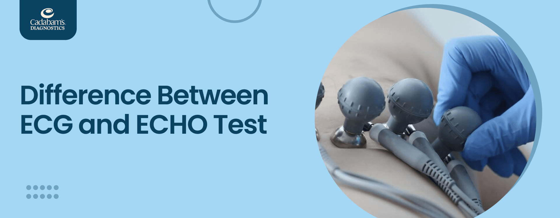
An ECG (electrocardiogram) is a medical test that measures the electrical activity of the heart over a period of time. It is a noninvasive, painless procedure that involves attaching electrodes to the skin, typically on the chest, arms, and legs, to detect the electrical impulses generated by the heart as it beats.
The ECG records these impulses and displays them as a series of waves and intervals on a graph, which helps doctors assess the heart’s rhythm, rate, and overall electrical function. ECG test results are commonly used to diagnose various heart conditions, such as arrhythmias, heart attacks, and other heart abnormalities.
Understanding Normal ECG Readings
An ECG (electrocardiogram) provides a visual representation of the electrical activity of the heart, helping to diagnose various heart conditions. Understanding the components of a normal ECG reading is essential for interpreting results accurately.
What Does a Normal ECG Report Look Like, and Its Components?
A normal ECG is a graphical representation of the electrical activity of the heart, showing specific patterns that indicate a healthy heart function. Key components of a normal ECG include heart rate, rhythm, waveform, and intervals.
Heart Rate
A normal heart rate on an ECG ranges from 60 to 100 beats per minute. The heart rate is measured by the distance between successive R waves (the peaks of the QRS complexes), which typically falls within the normal range of the ECG test.
Rhythm
A normal rhythm on an ECG is regular, with evenly spaced R-R intervals. The rhythm originates from the sinoatrial (SA) node, the heart’s natural pacemaker, leading to consistent and orderly heartbeats.
Waveform
The normal waveform on an ECG includes:
- P wave: Represents atrial depolarisation and precedes the QRS complex.
- QRS complex: This represents ventricular depolarisation and is the most prominent part of the ECG. It follows the P wave and has a sharp, tall appearance.
- T wave: Represents ventricular repolarisation and follows the QRS complex. It is usually a smooth, upward curve.
Intervals
Normal intervals on an ECG include:
- PR interval: Measures the time from the onset of the P wave to the start of the QRS complex, typically ranging from 120 to 200 milliseconds (ms).
- QRS duration: Measures the time taken for ventricular depolarisation, typically less than 120 ms.
- QT interval: Measures the time from the start of the Q wave to the end of the T wave, varying with heart rate but generally within 350 to 450 ms for men and 360 to 460 ms for women.
What is an Abnormal ECG Report?
An abnormal ECG report indicates deviations from the heart’s regular electrical activity, which can signify various cardiac conditions. Understanding the elements of an abnormal ECG is important for diagnosing and managing heart health issues.
Causes of Abnormal ECG Readings
Abnormal ECG test results can result from underlying heart conditions and other health issues. Understanding these causes helps in diagnosing and managing heart health effectively. Here are some common causes of abnormal ECG test results:
- Coronary Artery Disease (CAD): Coronary artery disease (CAD) happens when plaque buildup causes the coronary arteries, which provide blood to the heart muscle, to become narrowed or blocked. This condition can reduce blood flow to the heart, leading to chest pain (angina), shortness of breath, and, in severe cases, heart attacks. Early diagnosis and correct treatment are crucial to prevent further complications.
- Arrhythmias: Arrhythmias are irregular heartbeats caused by problems with the heart’s electrical system. They can range from harmless irregular beats to more serious conditions like atrial fibrillation or ventricular tachycardia, which may require medical intervention. Some arrhythmias can cause symptoms such as dizziness, palpitations, or fainting.
- Electrolyte Imbalances: Electrolytes such as potassium, calcium, and magnesium are crucial for normal heart function. Imbalances can disrupt the heart’s electrical activity, leading to abnormal heart rhythms or even cardiac arrest. These imbalances can be caused by dehydration, medications, or underlying health conditions and require prompt correction to restore normal heart function.
- Structural Heart Problems: Structural abnormalities in the heart, such as hypertrophy or congenital defects, can affect its electrical activity. These structural changes may lead to inefficient heart pumping or irregular rhythms, increasing the risk of heart failure. Treatment often involves medications, surgery, or procedures like ablation to correct these issues and restore normal function.
Abnormal Heart Rate in ECG Test Results
An abnormal heart rate refers to a heart rate that is either too fast (tachycardia) or too slow (bradycardia). Tachycardia is typically defined as a heart rate over 100 beats per minute, while bradycardia is a heart rate under 60 beats per minute.
An abnormal heart rate may be indicated in the ECG test results, which can be due to factors like stress, medications, or underlying heart conditions. A medical evaluation may be necessary to understand the cause and appropriate treatment.
Abnormal Heart Rhythm
An abnormal heart rhythm, or arrhythmia, means the heart beats irregularly. This can include conditions such as atrial fibrillation, where the heart beats irregularly and often rapidly, or ventricular fibrillation, a severe and life-threatening condition where the heart quivers instead of pumping effectively.
Arrhythmias can lead to symptoms like palpitations, dizziness, and fatigue and may require treatments ranging from medications to more invasive procedures like pacemaker implantation.
Abnormal Waveform
An abnormal waveform on an ECG can indicate issues with how the electrical signals are passing through the heart. This might include abnormal shapes or durations of the P wave, QRS complex, or T wave, which could suggest various cardiac conditions like hypertrophy, ischemia, or infarction.
Recognising abnormal waveforms in ECG test results is essential for diagnosing heart damage, and further diagnostic tests or treatments may be necessary depending on the specific condition.
Abnormal Intervals
Abnormal intervals refer to the timing between the different parts of the heart’s electrical cycle being outside the normal range. For example, a prolonged QT interval can increase the risk of sudden cardiac arrest, while a short PR interval may indicate pre-excitation syndromes such as Wolff-Parkinson-White syndrome.
Monitoring and addressing abnormal intervals are important for preventing potentially dangerous heart conditions and ensuring proper electrical conduction within the heart.
How to Prepare for an ECG Test?
Preparing for an ECG (electrocardiogram) test involves a few simple steps to ensure accurate results and a smooth testing process.
Before the Test
Avoid Caffeine and Smoking: Refrain from consuming caffeine or smoking for a few hours before the test, as these can affect your heart rate.
Wear Comfortable Clothing: Choose loose, comfortable clothing that can be easily removed or adjusted to attach the electrodes to your chest, arms, and legs.
Inform Your Doctor: Tell your doctor about any medications you are taking, as some drugs can influence the ECG results.
Follow Fasting Instructions: If your doctor has given specific instructions on how to fast before the test, follow them carefully.
Relax: Try to stay calm and relaxed before the test, as anxiety can affect your heart rate and potentially the results.
During the Test
Positioning: You will be asked to lie down on an examination table. The technician will attach electrodes to your chest, arms, and legs. These electrodes are connected to the ECG machine to record the electrical activity of your heart.
Stay Still: It’s important to lie still and breathe normally during the test to avoid any interference with the ECG recording. Movement can create artefacts that may affect the accuracy of the results.
Follow Instructions: The technician may ask you to hold your breath for a few seconds or remain still during certain parts of the test. Follow these instructions carefully.
Duration: The test typically takes about 5-10 minutes to complete, including the time to attach and remove the electrodes.
No Discomfort: An ECG is a painless procedure, and you should not feel any discomfort. If you experience any discomfort or have concerns, inform the technician immediately.
When to Consult a Doctor?
Consulting a doctor is crucial if you experience symptoms such as chest pain, shortness of breath, dizziness, or palpitations. Additionally, if your ECG test results or ECG normal report reveal any irregularities or if you have risk factors like a family history of heart disease, high blood pressure, diabetes, or high cholesterol, regular check-ups are recommended to monitor your heart health.
Interpreting Your ECG Results
Interpreting ECG results involves understanding the electrical activity of your heart. Abnormal results may indicate various conditions, such as arrhythmias, heart blockages, or previous heart attacks. It’s essential to have a healthcare professional explain your ECG test results to determine if further testing or treatment is needed.
Follow-up Steps After an Abnormal ECG
After an abnormal ECG, follow-up steps typically include additional diagnostic tests, such as an echocardiogram, stress test, or angiogram, to get a clearer picture of your heart’s condition. Your doctor may also recommend lifestyle changes, medications, or specific treatments based on the findings to address any underlying heart issues and improve your heart health.
Embracing a Heart-Healthy Lifestyle with Cadabam’s
Cadabam’s Diagnostics is a leading diagnostic centre that provides specialised services aimed at promoting heart health and overall wellness. Located across multiple locations in Bangalore, we offer a wide range of diagnostic tests, including comprehensive ECG screenings, radiology services, and laboratory investigations, all tailored to help you maintain a healthy heart.
Navigating heart health with Cadabam’s Diagnostics means receiving personalised care, expert medical advice, and continuous support. Our team of specialists is dedicated to clear communication, supporting you through each step from diagnosis to treatment, while equipping you with the information needed to make informed choices about your heart health.
At Cadabam’s, we believe that focusing on heart health not only lowers the risk of cardiovascular diseases but also boosts energy levels and enhances overall quality of life. For more information on heart health or to schedule a heart screening, visit our website or reach out to us at info@cadabamsdiagnostics.com. Let us partner with you in your journey towards a healthier, heart-conscious life.
FAQs
1. What is an ECG, and why is it important?
An ECG (electrocardiogram) is a test that measures the heart’s electrical activity through electrodes placed on the skin. It helps detect heart conditions like arrhythmias, heart attacks, and other abnormalities by displaying the heart’s rhythm and electrical impulses. Early diagnosis through ECGs enables timely treatment and better management of heart health.
2. What does a normal ECG reading look like?
A normal ECG test result shows a heart rate of 60-100 beats per minute with a consistent rhythm and includes distinct components like the P wave, QRS complex, and T wave. These represent different phases of heart function, such as atrial and ventricular depolarisation and repolarisation. Normal intervals between waves, such as the PR interval and QT interval, ensure proper heart function.
3. What causes abnormal ECG test results?
Abnormal ECG test results can result from various issues, such as coronary artery disease (CAD), where plaque buildup restricts blood flow, or arrhythmias, where the heart’s electrical signals are irregular. Other causes include electrolyte imbalances, structural heart defects, or abnormal heart rates, all of which affect the heart’s electrical activity and may require medical attention.
4. How should I prepare for an ECG test?
Before an ECG, it’s best to avoid caffeine or smoking, as these can elevate your heart rate and affect results. Wear loose, comfortable clothing to make attaching the electrodes easier, and inform your doctor about any medications you’re taking. Staying calm and following instructions during the test ensures accurate results.
5. What should I do after receiving an abnormal ECG test result?
After an abnormal ECG test result, your doctor may suggest additional tests, such as an echocardiogram or stress test, to get a clearer picture of your heart’s condition. Treatment could involve lifestyle changes, medications, or more specific procedures, depending on the diagnosis. Regular monitoring and follow-up care are essential for managing your heart health effectively.
6. How to check if an ECG report is normal?
To check if an ECG report is normal, look for specific indicators, including a heart rate between 60 and 100 beats per minute, a regular rhythm, and standard intervals, such as the PR interval (120-200 milliseconds) and the QRS duration (less than 120 milliseconds). Normal waveforms should include clearly identifiable P waves, QRS complexes, and T waves without any unusual peaks or irregularities. If unsure, it’s always best to consult a healthcare professional to ensure accurate interpretation of the report.
7. How to read an ECG report for accurate results?
Learning how to read an ECG report involves understanding the various waves, intervals, and rhythms that represent the heart’s electrical activity. The P wave indicates atrial depolarisation, the QRS complex shows ventricular depolarisation, and the T wave represents ventricular repolarisation. Observing these components in relation to each other and comparing them to standard values can help identify whether the ECG reflects normal heart function. For a more accurate reading, review the heart rate, rhythm, and intervals, or consult a trained professional.