ECG vs. Echocardiogram: A Guide to Heart Diagnostics
Verified by: Dr. Shreyas Cadabam
When it comes to heart diagnostics, understanding the differences between an Electrocardiogram (ECG) and an Echocardiogram is crucial for effective healthcare. These two tests play vital roles in detecting and managing heart conditions, offering unique insights into heart health.
At Cadabam's Diagnostics, we prioritise your well-being by providing comprehensive diagnostic services, including ECG and Echocardiogram, ensuring you receive accurate and timely results for optimal heart care.
Introduction to Heart Health Monitoring
Monitoring heart health is essential for early detection and effective management of cardiovascular conditions. Regular heart health check-ups can help identify potential issues before they become serious, ensuring a proactive approach to wellness.
What is an ECG?
An Electrocardiogram (ECG) is a non-invasive test that records the heart's electrical activity over time. It is a fundamental tool in diagnosing various heart conditions, such as arrhythmias, heart attacks, and other cardiac abnormalities.
An ECG analyses the heart's electrical signals to offer valuable insights into its rhythm, structure, and function.\\\\\\\
How does an ECG work?
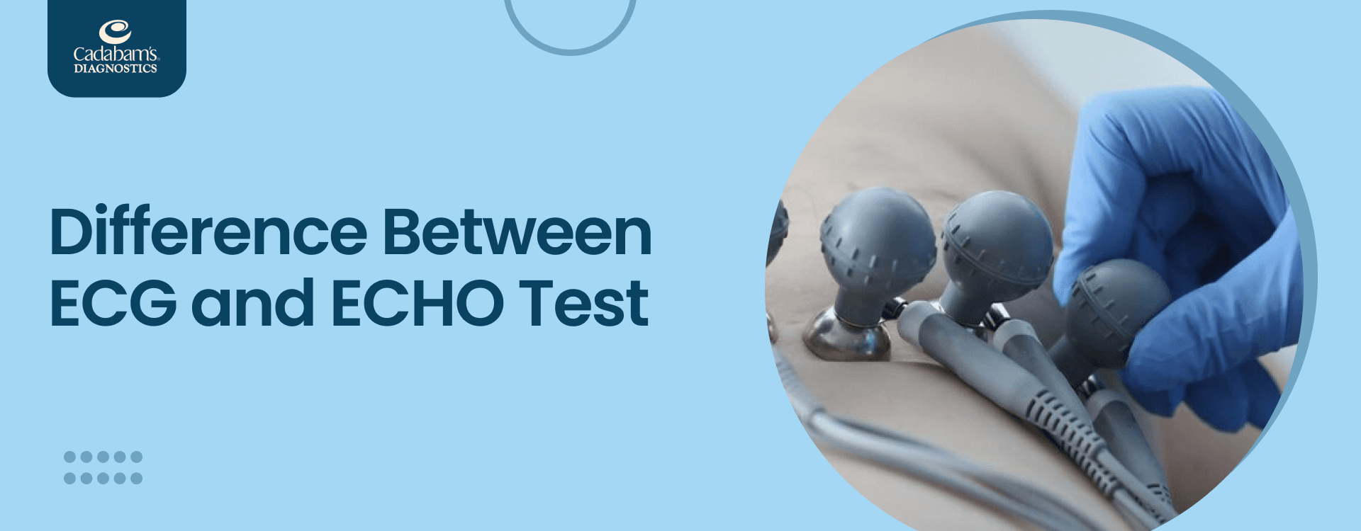
An ECG involves placing small adhesive electrodes on the skin of the chest, arms, and legs. These electrodes are linked to an ECG machine that detects the heart's electrical impulses.
The machine then records these impulses as waves on a graph, which a healthcare professional can interpret.
What is an Echo (Echocardiogram)?
An Echocardiogram, often called an Echo, is a non-invasive diagnostic test that uses ultrasound technology to produce detailed images of the heart.
This test visually represents the heart's structure and function, allowing healthcare providers to assess the heart's chambers, valves, and blood flow.
How Ultrasound Technology Maps the Heart?
Ultrasound technology in an Echocardiogram uses high-frequency sound waves to produce images of the heart. A device known as a transducer is positioned on the chest, sending out sound waves that reflect off the heart's structures.
These echoes are captured and converted into moving images displayed on a monitor. This process allows for real-time visualisation of the heart, enabling a detailed assessment of its anatomy and functionality.
Echocardiogram Applications in Heart Health
- Diagnosing Heart Conditions: Detects heart valve disorders, cardiomyopathy, congenital heart defects, and pericardial disease.
- Evaluating Heart Function: This assesses the heart's pumping efficiency and blood flow, especially after a heart attack or in heart failure cases.
- Guiding Treatment Decisions: Aids in planning surgeries, evaluating treatment effectiveness, and monitoring heart condition progress.
- Detecting Blood Clots and Tumours: Identifies abnormal masses like blood clots or tumours within the heart.
- Assessing Heart Damage: Evaluate the extent of damage from conditions like heart attacks.
When do you need ECGs and ECHO for Heart Health Monitoring?
Both ECGs and Echo tests are essential tools for heart health monitoring, and their use depends on specific symptoms and medical needs.
ECG
You may need an ECG if you experience:
- Chest Pain: To check for heart attack or angina.
- Palpitations: To detect abnormal heart rhythms (arrhythmias).
- Shortness of Breath: To evaluate heart function and detect heart failure.
- Dizziness or Fainting: To identify potential heart-related causes.
- High Blood Pressure: To assess the heart’s response to hypertension.
- Routine Check-Up: For baseline heart health monitoring, especially if you have a family history of heart disease.
Echo
You may need an Echo if you experience:
- Heart Murmurs: To investigate abnormal heart sounds.
- Swelling in the Legs (Edema): To evaluate heart function and detect heart failure.
- Shortness of Breath: To assess heart structure and function.
- Unexplained Fatigue: To check for underlying heart conditions.
- Known Heart Disease: To monitor existing conditions such as cardiomyopathy or valvular heart disease.
- Post-Heart Attack: To evaluate the extent of heart damage and guide treatment.
How are ECG and Echocardiogram Procedures Conducted?
An ECG is conducted by placing small electrodes on the chest, arms, and legs to record the heart's electrical activity. It typically takes a few minutes. An Echocardiogram involves using a handheld device called a transducer to emit ultrasound waves and create moving images of the heart. It usually takes 30 to 60 minutes.
ECG Procedures
Time
The ECG procedure typically takes a few minutes to complete.
Procedures
- Small adhesive electrodes are placed on the chest, arms, and legs.
- The electrodes are connected to an ECG machine that records the heart's electrical activity.
- The patient remains still while the machine captures the electrical signals and displays them as waves on a graph for analysis.
Echo Procedures
Time
An Echocardiogram usually takes around 30 to 60 minutes to complete.
Procedures
- A technician applies a special gel to the chest to enhance sound wave transmission.
- A handheld device called a transducer is moved over the chest area, emitting ultrasound waves.
- The sound waves bounce off the heart structures and are captured by the transducer, creating real-time moving images displayed on a monitor for detailed analysis.
ECG vs Echocardiogram: Interpreting Test Results
Understanding the results of these tests can provide a clearer picture of your heart's health and guide appropriate treatment plans. For accurate interpretation and personalised advice, always consult with a healthcare professional.
Interpreting ECG Readings
Understanding an ECG reading provides crucial insights into heart health. Below are key elements analysed during an ECG:
- Heart Rate:
- Normal: 60-100 beats per minute (bpm)
- Bradycardia: Less than 60 bpm
- Tachycardia: More than 100 bpm
- Heart Rhythm:
- Regular: Consistent intervals between beats
- Irregular: Variable intervals indicating potential arrhythmias
- P Wave:
- Represents atrial depolarisation
- Abnormalities may indicate atrial enlargement or arrhythmias
- QRS Complex:
- Represents ventricular depolarisation
- Normal duration: 0.06-0.10 seconds
- Prolonged QRS could suggest bundle branch block or ventricular hypertrophy
- ST Segment:
- It should be flat (isoelectric)
- Elevation or depression may indicate myocardial ischemia or infarction
- T Wave:
- Represents ventricular repolarisation
- Abnormalities may signal ischemia, electrolyte imbalances, or other heart conditions
- PR Interval:
- Duration: 0.12-0.20 seconds
- Prolonged intervals can indicate first-degree heart block
- QT Interval:
- Varies with heart rate but generally less than 0.44 seconds
- Prolonged QT increases the risk of arrhythmias
Common Findings:
- Normal Sinus Rhythm (NSR): Regular rhythm, normal heart rate, normal P wave, QRS complex, and T wave
- Atrial Fibrillation (AFib): Irregular rhythm, absence of distinct P waves
- Myocardial Infarction (MI): ST elevation or depression, presence of pathologic Q waves
Analysing Echo Results
An Echocardiogram offers a detailed view of the heart's structure and function. Below are key aspects evaluated during an Echo:
- Heart Chambers:
- Examines the size, shape, and thickness of the heart's chambers
- Detects abnormalities like chamber enlargement or structural defects
- Heart Valves:
- Assesses the function and structure of heart valves
- Detects conditions like stenosis (narrowing) or regurgitation (leakage)
- Ejection Fraction (EF):
- Measures the percentage of blood pumped out by the left ventricle with each contractio
- Used to evaluate the heart's pumping efficiency
- Wall Motion:
- Evaluates the movement of the heart walls
- Identifies areas of weakened or damaged heart muscle, which may suggest previous heart attacks
- Blood Flow (Doppler Ultrasound):
- Assesses the flow of blood through the heart and major vessels
- Detects abnormal flow patterns or blockages
- Pericardium:
- Examines the sac surrounding the heart (pericardium)
- Detects inflammation, fluid buildup, or other abnormalities in the pericardium
Common Findings:
- Normal Heart Function: Properly sized chambers, normal valve function, and efficient ejection fraction
- Valve Disorders: Detection of stenosis or regurgitation affecting the heart’s ability to pump blood effectively
- Cardiomyopathy: Enlarged heart chambers or abnormal heart muscle thickness, impacting heart function
- Heart Failure: Low ejection fraction and abnormal wall motion indicating impaired pumping ability
- Blood Clots/Tumours: Presence of abnormal masses or clots in the heart
Embracing Cardiac Health with Cadabam’s Diagnostics
At Cadabam’s Diagnostics, we are committed to providing top-tier diagnostic services for heart health. Specialising in both ECG and Echocardiogram tests, we offer comprehensive solutions to monitor, detect, and manage various cardiac conditions. Our team of experts ensures thorough preparation, accurate diagnostics, and personalised care to help you navigate your heart health journey with confidence.
With Cadabam’s, you’ll experience clear communication, professional guidance, and access to the latest diagnostic technology, ensuring that every step of your heart health monitoring is seamless and informed. Whether you're undergoing routine check-ups or need advanced cardiac diagnostics, our multidisciplinary team is here to support you every step of the way.
For more information or to schedule your ECG or Echo test, visit our website or reach out to us at info@cadabamsdiagnostics.com. Let us be your trusted partner in cardiac care, delivering the highest standards of medical excellence and compassionate support.