Scrotum Ultrasound: Diagnostic Uses and Procedure
Verified by: Dr. Shreyas Cadabam
"
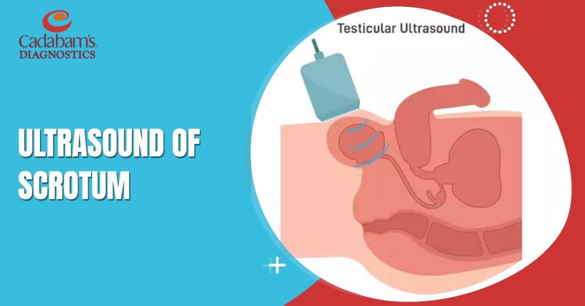
Ultrasound of the scrotum, also known as scrotal ultrasound, is a diagnostic imaging technique that uses high-frequency sound waves to create detailed images of the scrotum and its contents, including the testicles, epididymis, and surrounding structures. Scrotal ultrasound can sometimes be an emergency in cases of acute pain in order to determine if there is any torsion that can be reversed. It is also used as an important tool to assess size, parenchymal abnormalities and other features in cases of infertility.
Here’s how scrotal ultrasound works and its applications:
Varicocele
Varicocele is an unusual dilation of the veins above the testicles and can cause secondary infertility because of reduced sperm count and motility. An ultrasound of the spectrum shows tangles of the spermatic cord that are twisted into tufts.
Epididymal cyst
This is the blockage of your vas deferens in the epididymis. It can lead to male infertility, if not treated early.
Testicular torsion
This is a dangerous disease that, if not promptly treated, may lead to testicular necrosis and testicular atrophy. Ultrasound of the testicles and scrotum helps in detecting the degree of torsion so that they can do timely treatment.
Epididymitis
This results from retrograde infection from the prostate gland and bladder.
Orchitis
Usually, orchitis occurs due to acute infection of the testicles, invaders, streptococcus, and staphylococcus. The symptoms are painful, like pain in the testicle, heaviness in the scrotum, and swelling of the testicle. An ultrasound image will detect swelling and inflammation.
Scrotal trauma:
Scrotal ultrasound is useful in assessing traumatic injuries to the scrotum, such as testicular rupture, hematomas, or other internal damage. It can help determine the extent of the injury and guide appropriate management.
Procedure:
During a scrotal ultrasound, a gel is applied to the scrotum, and a handheld transducer is moved over the area to capture images. The procedure is generally painless and non-invasive. The technician or radiologist will evaluate the scrotal structures, including the testicles, epididymis, and blood vessels, to assess their size, shape, and blood flow patterns.
The radiologist based on the images can identify the pathology in the organs based on the size, echopattern and vascularity. If you have scrotal pain, swelling, or concerns about any scrotal-related issues, it is recommended to visit a radiologist who could identify/ rule out any pathologies.
Find the Best Ultrasound clinic in Bangalore for your scrotal ultrasound.