No. You only hear soft knocking sounds; earplugs are provided.
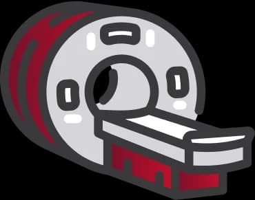
MRI
MRI Brain Csf Flow Study
Centre visitAdvanced equipment
Senior
Flat 10% off for senior citizens
Family
Add a family member for 20% discount
60 Mins
1M
4.9
5
MRI BRAIN CSF FLOW STUDY Overview
List of Parameters
Parameters Considered During CSF Flow Study
When to Take Test
When to Take Test
Benefits
Benefits of MRI CSF Flow Study
Diagnosed Conditions by CSF Flow Study Radiology
Preparing for Test
Preparation for MRI Brain CSF Flow Study
Risks & Limitations in MRI CSF Flow Study
Test Results
Results and Interpretations of the MRI Brain CSF Flow Study
| Finding / Observation | Description (Example Values/Observations) | General Interpretation/Significance (Examples) |
|---|---|---|
| Normal flow | Smooth, forward-moving CSF | No obstruction |
| Reduced velocity | <2 cm/s peak systolic | Possible aqueductal stenosis |
| Reversed flow | CSF moves backward | Chiari malformation sign |
| High stroke volume | >42 µL/heartbeat | Communicating hydrocephalus |
| Turbulent jets | Chaotic flow pattern | CSF leak or fistula |
| Patency of CSF Pathways | Aqueduct of Sylvius appears patent; fourth ventricular outlets unremarkable | No evidence of obstruction; normal CSF circulation expected. |
| Patency of CSF Pathways | Significant narrowing noted at the aqueduct level | Suggestive of aqueductal stenosis, potentially causing obstructive hydrocephalus. |
| Shunt Patency (if VP shunt present) | Visible pulsatile flow within ventricular catheter and shunt tubing | Shunt appears patent and functional. |
| Shunt Patency (if VP shunt present) | No flow signal detected within shunt tubing; ventricles enlarged | Possible shunt malfunction or blockage; clinical correlation needed. |
| Presence of Abnormal Flow Jets | Abnormal turbulent jetting flow from third ventricle into aqueduct | May indicate pressure gradient or partial obstruction c |
| Qualitative CSF Pulsatility | Generally diminished CSF pulsatility throughout supratentorial cisterns | Seen in intracranial hypotension or advanced hydrocephalus with poor compliance. |